Experimental objects: fungi
|
|
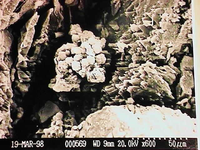
1. SEM photo of fungal microcolony on the surface of marble. |
|
|
|
|
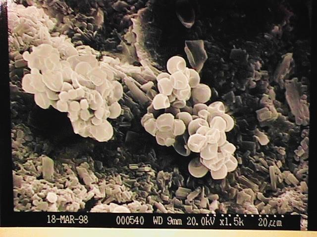
2. Experimentally obtained fungal microcolonies on the surface of calcium gluconate tablets supplied with CuSO4. Growth in the yeast form. |
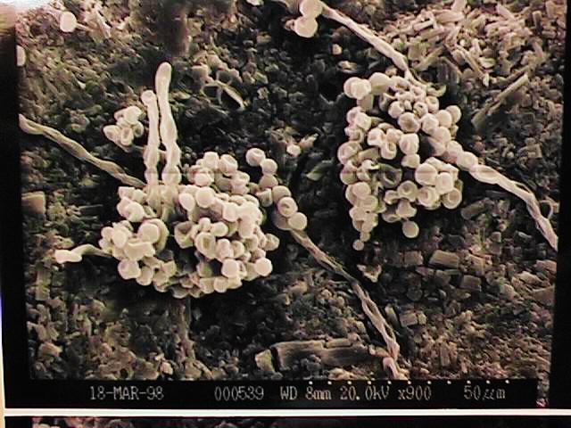
3. Experimentally obtained fungal microcolonies on the surface of calcium gluconate tablets without any additions. Mycelial growth. |
 |
4. Phaeococcomyces chersonesos Strain Ch 49. Yeast form. |
 |
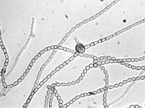
5. Phaeococcomyces chersonesos Strain Ch 49. Mycelial form. |
 |
6. Ph. chersonesos (Ch 49). Mixed culture with pseudomycelium and yeast. |
 |
7. Ph. chersonesos (Ch 49). Chlamydospores in yeast culture. |
 |
8. Ph. chersonesos (Ch 49). Proliferating intercalary chlamydospore on the mycelium. |
 |
9. Ph. chersonesos (Ch 49). Yeast to mycelium transition under the action of isopropyl alcohol. 1- initial yeast cells; 2 - secondary cells of a greater size (up to 5 times greater); 3 - pseudomycelial and further mycelial growth. Bar = 20 mkm. |